Natsu Natsu
Age-Specific Analysis of Cortical and Trabecular Bone Mineral Density in the Femur and Lumbar Spine Using a Large-Scale CT Database大規模CTデータベースを用いた大腿骨および腰椎における皮質骨・海綿骨密度の年代別解析
This project investigates age-related changes in cortical and trabecular bone mineral density (BMD) in the femur and lumbar spine, using a large-scale clinical CT database and advanced image analysis methods. Bone mineral density is a well-established predictor of fracture risk, and its assessment is central to the diagnosis and management of osteoporosis, a major public health concern affecting hundreds of millions of people worldwide, particularly postmenopausal women and elderly men. However, conventional BMD measurement using dual-energy X-ray absorptiometry (DXA) has important limitations: as a two-dimensional projection technique, DXA averages the bone density over a projected area without distinguishing between cortical and trabecular compartments or capturing spatial distribution patterns. These distinctions are clinically relevant, as cortical and trabecular bone have different structural roles, respond differently to aging and hormonal changes, and are associated with different fracture mechanisms.
本研究は、大規模臨床CTデータベースと高度な画像解析手法を用いて、大腿骨および腰椎における皮質骨と海綿骨の骨塩量(BMD)の年代別変化を調査します。BMDは骨折リスクの確立した予測因子であり、骨粗鬆症の診断と管理において中心的な役割を担います。しかし、DXAによる従来のBMD計測には重要な限界があります。二次元投影技術であるDXAは、皮質骨と海綿骨を区別したり空間分布パターンを捉えたりすることなく投影面積上で平均化してしまいます。皮質骨と海綿骨は構造的役割・加齢・ホルモン変化への反応・骨折メカニズムが異なるため、この区別は臨床的に重要です。
Focus研究焦点
Age-Specific Analysis of Cortical and Trabecular Bone Mineral Density in the Femur and Lumbar Spine Using a Large-Scale CT Database大規模CTデータベースを用いた大腿骨および腰椎における皮質骨・海綿骨密度の年代別解析
This project investigates age-related changes in cortical and trabecular bone mineral density (BMD) in the femur and lumbar spine, using a large-scale clinical CT database and advanced image analysis methods. Bone mineral density is a well-established predictor of fracture risk, and its assessment is central to the diagnosis and management of osteoporosis, a major public health concern affecting hundreds of millions of people worldwide, particularly postmenopausal women and elderly men. However, conventional BMD measurement using dual-energy X-ray absorptiometry (DXA) has important limitations: as a two-dimensional projection technique, DXA averages the bone density over a projected area without distinguishing between cortical and trabecular compartments or capturing spatial distribution patterns. These distinctions are clinically relevant, as cortical and trabecular bone have different structural roles, respond differently to aging and hormonal changes, and are associated with different fracture mechanisms.
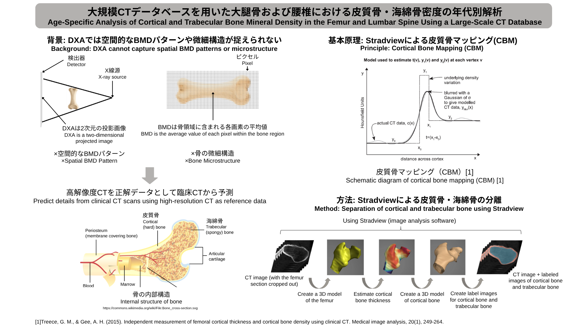
The proposed approach addresses these limitations by directly analyzing CT images, which provide full three-dimensional volumetric information about bone structure. The key methodological tool is Cortical Bone Mapping (CBM) as implemented in the Stradview software. CBM is a technique that models the CT intensity profile across the cortical surface at each vertex of a 3D bone model, fitting a parameterized model that estimates cortical thickness, cortical density, and trabecular density simultaneously at each location. This approach allows the spatial distribution of bone density to be mapped across the entire surface of the femur or vertebra, rather than being summarized by a single average value as in DXA.
The analysis pipeline begins with the segmentation of CT images to separate the full bone mask into its cortical and trabecular components. This segmentation is performed using Stradview, which produces three types of labeled images for each CT scan: the whole bone mask, the cortical bone label, and the trabecular bone label. These labels are used to extract region-specific density measurements that reflect the distinct mechanical properties and biological behaviors of the two bone compartments. The dataset used in this study includes 110 subjects spanning a broad age range from 50 to over 90 years, with a predominantly female composition reflecting the epidemiology of osteoporosis.
The study examines age-related trends in both compartments and both anatomical sites, generating profiles of how cortical and trabecular BMD change across different decades of life. A particular focus is placed on the differential rate of bone loss: trabecular bone, which has a higher metabolic turnover rate due to its highly vascularized spongy structure, is expected to show more pronounced and earlier density reductions compared to cortical bone, especially in women following menopause. The results confirm this expectation, showing that trabecular bone density declines more steeply with age, particularly in females.
An additional analysis investigates the correlation between BMD measured at the femur and at the lumbar spine. The results reveal that trabecular bone density shows a stronger inter-site correlation than cortical bone density, which is attributed to the systemic nature of trabecular bone remodeling — driven by hormonal and metabolic factors that affect the whole skeleton — versus the more locally influenced remodeling of cortical bone, which is additionally regulated by mechanical loading patterns specific to each anatomical site. These findings contribute to a more nuanced, spatially resolved understanding of skeletal aging and may inform the development of CT-based BMD biomarkers for improved osteoporosis risk stratification.
本研究は、大規模臨床CTデータベースと高度な画像解析手法を用いて、大腿骨および腰椎における皮質骨と海綿骨の骨塩量(BMD)の年代別変化を調査します。BMDは骨折リスクの確立した予測因子であり、骨粗鬆症の診断と管理において中心的な役割を担います。しかし、DXAによる従来のBMD計測には重要な限界があります。二次元投影技術であるDXAは、皮質骨と海綿骨を区別したり空間分布パターンを捉えたりすることなく投影面積上で平均化してしまいます。皮質骨と海綿骨は構造的役割・加齢・ホルモン変化への反応・骨折メカニズムが異なるため、この区別は臨床的に重要です。
提案手法は、骨構造の完全な三次元体積情報を提供するCT画像を直接解析することでこれらの限界に対処します。主要な方法論的ツールはStradviewソフトウェアで実装された皮質骨マッピング(CBM)です。CBMは三次元骨モデルの各頂点での皮質表面横断方向のCT輝度プロファイルをモデル化し、パラメータ化モデルを当てはめることで各位置の皮質厚・皮質密度・海綿骨密度を同時推定します。これにより、DXAのような単一平均値ではなく大腿骨や椎体の全表面にわたる空間的な骨密度分布マップが得られます。
解析パイプラインはCT画像のセグメンテーションから始まり、全骨マスクを皮質骨成分と海綿骨成分に分離します。Stradviewが各CTスキャンに対して全骨マスク・皮質骨ラベル・海綿骨ラベルの三種のラベル画像を生成し、各骨区画の異なる機械的特性と生物学的挙動を反映する領域特異的密度計測を可能にします。本研究のデータセットには50歳から90歳超の広い年齢範囲にわたる110名が含まれ、骨粗鬆症の疫学を反映して女性が多数を占めています。
研究では両骨区画・両解剖学的部位の年代別トレンドを調査し、皮質骨と海綿骨のBMDが人生の各年代でどのように変化するかをプロファイル化します。特に骨量減少率の差異に着目しています。代謝回転率が高い海綿骨は皮質骨より顕著で早期の密度低下を示すことが予想され、特に閉経後の女性でその傾向が顕著です。結果はこの予測を確認し、海綿骨密度が年齢とともにより急峻に低下することを示しています。
大腿骨と腰椎のBMD相関の追加解析では、海綿骨密度が皮質骨密度よりも部位間相関が強いことが明らかになりました。これは、全身に影響するホルモン・代謝因子に駆動される海綿骨リモデリングの全身的性質と、各解剖学的部位特有の力学的負荷パターンによる影響を受ける皮質骨リモデリングのより局所的な性質に起因すると考えられます。これらの知見は骨格老化の空間的に解像されたより詳細な理解に貢献し、骨粗鬆症リスク層別化の改善のためのCTベースのBMDバイオマーカー開発に情報を提供します。
Project 8
Predicting Internal Body Structures from 3D Surface Scans
3D体表面から身体内部の構造を予測
Statistical Shape Model
Surface-to-Bone Estimation
3D Reconstruction
Non-invasive Imaging
Musculoskeletal Modeling