Research研究
Research categories and active projects.研究カテゴリと進行中の研究。
Browse the lab by research category, then dive into individual projects and studies.
研究カテゴリで全体像を見渡し、その先にある個別研究へ進めます。
Projects個別研究
Filtered research projects. カテゴリで絞り込まれた研究一覧。

Now
Motion Analysis of Ballet Movementsクラシックバレエにおける個別筋骨格解析を活用した動作解析
This project addresses the challenge of quantitatively evaluating ballet posture and movement quality through computational methods grounded in subject-specific musculoskeletal simulation. Classical ballet is a highly technical art form where the quality of posture — including joint angles, muscle coordination, and limb alignment — is traditionally assessed by trained experts according to established aesthetic and functional criteria. However, such expert evaluation is inherently subjective, time-consuming, and difficult to scale. The long-term goal of this research is to develop an objective, automated assessment system capable of producing posture evaluation scores that are comparable in quality to those given by domain experts, thereby supporting dancer training and reducing injury risk.
本研究は、個別筋骨格シミュレーションに基づく計算手法を用いて、バレエの姿勢と動作の質を定量的に評価することを目的としています。クラシックバレエは、関節角度・筋肉の協調動作・四肢のアライメントなど、姿勢の質が伝統的に専門家による評価に委ねられてきた高度に技術的な芸術形式です。しかしながら、専門家による評価は本質的に主観的であり、時間がかかるうえ、スケールアップが困難です。本研究の長期的な目標は、専門家と同等の精度で姿勢評価スコアを自動生成できる客観的システムを開発し、ダンサーのトレーニング支援と負傷リスクの軽減を実現することです。
Now
Predicting Survival in Fulminant Myocarditis Using a Large-Scale Pathology Foundation Model大規模病理基盤モデルを用いた劇症型心筋炎の生存予測
This project addresses the problem of prognosis prediction in fulminant myocarditis, a rare and severe form of myocarditis characterized by acute inflammatory infiltration of the cardiac muscle that leads to rapid deterioration of cardiac function. Due to its acute and life-threatening nature, fulminant myocarditis requires urgent clinical management, and early identification of patients at high risk of mortality is essential for guiding treatment decisions. Despite the clinical urgency, conventional computational models based on convolutional neural networks applied to histopathological images have consistently struggled to achieve high predictive accuracy, partly due to the limited availability of annotated data and the complex visual patterns associated with the disease.
本研究は劇症型心筋炎の予後予測を対象としています。劇症型心筋炎は心筋への急性炎症性浸潤によって心機能が急速に低下する稀かつ重篤な疾患であり、死亡リスクが高い患者の早期同定は治療戦略の決定に不可欠です。しかし、病理組織画像にCNNを適用した従来の計算モデルは、アノテーションデータの少なさや疾患に特有の複雑な視覚的パターンにより、高い予測精度の達成に苦戦してきました。

Now
Spatiotemporal Analysis of Swallowing Movement via 4DCT4DCTを用いた嚥下運動の時空間分析
This project focuses on the spatiotemporal analysis of swallowing mechanics using four-dimensional computed tomography (4DCT), a dynamic imaging modality that acquires volumetric CT data at multiple successive time points to capture the motion of anatomical structures over time. Swallowing is a highly coordinated neuromuscular process involving the simultaneous movement of dozens of structures including the tongue, hyoid bone, larynx, pharynx, and esophagus. Dysphagia — impaired swallowing — is a clinically important condition associated with aspiration pneumonia, malnutrition, and reduced quality of life, particularly in elderly and neurologically impaired populations. A precise, quantitative understanding of swallowing kinematics is therefore essential both for clinical diagnosis and for the development of targeted rehabilitation strategies.
本研究は、四次元CT(4DCT)を用いた嚥下動態の時空間分析に取り組んでいます。4DCTは連続した複数の時点で体積CTデータを取得する動態撮像モダリティであり、解剖学的構造の動きを経時的に捉えることができます。嚥下は、舌・舌骨・喉頭・咽頭・食道など多数の構造が関与する高度に協調されたの神経筋プロセスです。嚥下障害(嚥下困難)は誤嚥性肺炎・栄養不良・QOL低下と関連する臨床的に重要な状態であり、特に高齢者や神経障害患者に多く見られます。

Now
Development of an Automated Diagnostic System for Osteonecrosis of the Femoral Head Classification Based on MRI ImagesMRI画像から複数種類の病型分類に基づく大腿骨頭壊死症診断を行う自動システムの構築
This project targets the automated classification of osteonecrosis of the femoral head (ONFH), a debilitating orthopedic condition in which the blood supply to the femoral head is disrupted, leading to ischemic necrosis of the bone. ONFH can progress to femoral head collapse and secondary osteoarthritis if not treated at an early stage, making accurate severity assessment a critical component of clinical management. Providing optimal treatment — ranging from conservative management for mild stages to surgical interventions such as core decompression or total hip arthroplasty for advanced stages — depends fundamentally on an accurate and consistent classification of disease severity. However, current clinical practice faces a significant limitation: even among experienced clinicians, the classification of ONFH severity can be inconsistent, leading to variability in treatment decisions.
本研究は、大腿骨頭壊死症(ONFH)の自動分類システムの開発を目的としています。ONFHは大腿骨頭への血液供給が遮断され骨の虚血性壊死が生じる整形外科疾患であり、早期治療なしには大腿骨頭の圧潰と続発性変形性股関節症へと進行します。軽症への保存療法から進行例への骨頭温存手術・人工関節置換術まで、最適な治療の提供には正確かつ一貫した重症度分類が不可欠です。しかし現状では、経験豊富な臨床医の間でも重症度分類に一貫性が欠けるという課題があります。

Now
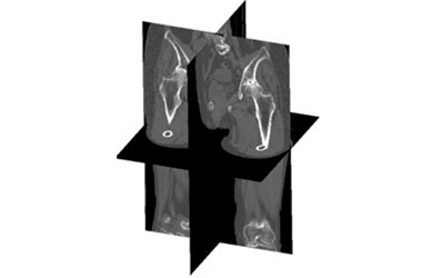
Age-Specific Analysis of Cortical and Trabecular Bone Mineral Density in the Femur and Lumbar Spine Using a Large-Scale CT Database大規模CTデータベースを用いた大腿骨および腰椎における皮質骨・海綿骨密度の年代別解析
This project investigates age-related changes in cortical and trabecular bone mineral density (BMD) in the femur and lumbar spine, using a large-scale clinical CT database and advanced image analysis methods. Bone mineral density is a well-established predictor of fracture risk, and its assessment is central to the diagnosis and management of osteoporosis, a major public health concern affecting hundreds of millions of people worldwide, particularly postmenopausal women and elderly men. However, conventional BMD measurement using dual-energy X-ray absorptiometry (DXA) has important limitations: as a two-dimensional projection technique, DXA averages the bone density over a projected area without distinguishing between cortical and trabecular compartments or capturing spatial distribution patterns. These distinctions are clinically relevant, as cortical and trabecular bone have different structural roles, respond differently to aging and hormonal changes, and are associated with different fracture mechanisms.
本研究は、大規模臨床CTデータベースと高度な画像解析手法を用いて、大腿骨および腰椎における皮質骨と海綿骨の骨塩量(BMD)の年代別変化を調査します。BMDは骨折リスクの確立した予測因子であり、骨粗鬆症の診断と管理において中心的な役割を担います。しかし、DXAによる従来のBMD計測には重要な限界があります。二次元投影技術であるDXAは、皮質骨と海綿骨を区別したり空間分布パターンを捉えたりすることなく投影面積上で平均化してしまいます。皮質骨と海綿骨は構造的役割・加齢・ホルモン変化への反応・骨折メカニズムが異なるため、この区別は臨床的に重要です。

Now
Sex and Age-related Analysis of Cortical and Trabecular Bone Density in the Femur and Lumbar Spine Using a Large-scale CT Database大規模CTデータベースを用いた大腿骨と腰椎における皮質骨・海綿骨密度の性別・年代別解析
This project extends the analysis of cortical and trabecular bone mineral density (BMD) to explicitly incorporate sex as a stratification variable, building on a dataset of 110 subjects aged between 50 and over 90 years with a female-dominant composition reflecting the clinical epidemiology of osteoporosis. The inclusion of both male and female subjects across multiple age decades enables a rigorous investigation of how biological sex modulates the trajectory of age-related bone loss in each of the two main bone compartments and at two clinically important skeletal sites — the proximal femur and the lumbar spine.
本研究は、50歳から90歳超にわたる110名(骨粗鬆症の臨床疫学を反映した女性多数構成)のデータセットを基に、皮質骨・海綿骨のBMD解析に性別を層別化変数として明示的に組み込んだ拡張研究です。複数の年代にわたる男女被験者の包含により、生物学的性別が各骨区画の年代別骨量減少軌跡にどのような影響を与えるかを厳密に調査することが可能となっています。