Research研究
Research categories and active projects.研究カテゴリと進行中の研究。
Browse the lab by research category, then dive into individual projects and studies.
研究カテゴリで全体像を見渡し、その先にある個別研究へ進めます。
Projects個別研究
Filtered research projects. カテゴリで絞り込まれた研究一覧。
Now
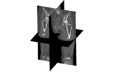
Pelvic Tilt Estimation Using Depth Maps from a Single Plain Radiograph一方向単純X線画像からの深度画像を用いた骨盤傾斜角推定
This project focuses on the estimation of pelvic tilt angles — sagittal, coronal, and axial — from a single anteroposterior plain radiograph, using a deep learning-based approach that relies on intermediate depth map representations. Pelvic tilt is a clinically critical parameter in orthopedic practice, particularly in the preoperative planning for total hip arthroplasty (THA). The orientation of the pelvis in three-dimensional space directly influences the functional positioning of the acetabular cup, and inaccurate tilt estimation can lead to suboptimal implant placement, contributing to complications such as dislocation, impingement, or accelerated wear. Currently, the gold standard for measuring pelvic tilt is three-dimensional CT imaging, which provides accurate volumetric data but involves higher radiation doses, greater cost, and longer acquisition times compared to plain radiography.
本研究は、深層学習に基づく深度マップ推定手法を用いて、単一の正面単純X線画像から骨盤の傾斜角(矢状面・冠状面・軸位面)を推定することを目的としています。骨盤傾斜は整形外科臨床において非常に重要なパラメータであり、特に人工股関節全置換術(THA)の術前計画において中心的な役割を果たします。骨盤の三次元的な向きは臼蓋カップの機能的位置決めに直接影響し、傾斜の推定誤差は脱臼・インピンジメント・摩耗促進などの合併症につながるリスクがあります。現在の標準的手法はCT撮影ですが、被ばく線量・費用・撮影時間の面で単純X線に劣ります。
Now
Anterior-Posterior Depth Estimation from a Single X-ray Image単一方向X線画像からの前後面深度推定
This project addresses the problem of recovering three-dimensional structural information from a single frontal X-ray image, with a specific focus on the vertebral column. While a single anteroposterior radiograph provides rich two-dimensional information about the size, shape, and alignment of vertebral bodies, it fundamentally lacks the depth dimension — the extent of each structure along the anterior-posterior (AP) axis, i.e., the axis perpendicular to the imaging plane. This missing dimension is critical for a complete understanding of spinal anatomy, particularly for assessing pathological conditions such as vertebral fractures, spondylolisthesis, or degenerative disc disease, where the three-dimensional geometry of the spine plays a central role in both diagnosis and treatment planning.
本研究は、単一の正面X線画像から脊椎の三次元構造情報を復元する問題に取り組んでいます。正面X線画像は椎体の大きさ・形状・アライメントに関する豊富な二次元情報を提供しますが、前後方向(AP方向、すなわち撮影面に垂直な軸)の奥行き次元が根本的に欠如しています。この欠損次元は脊椎解剖の完全な理解に不可欠であり、椎体骨折・脊椎すべり症・変性椎間板疾患などの病的状態の診断と治療計画において特に重要です。
Now
Predicting Internal Body Structures from 3D Surface Scans3D体表面から身体内部の構造を予測
This project addresses the problem of non-invasively estimating the three-dimensional geometry of internal skeletal structures from external body surface scans. In many clinical and biomechanical applications, knowledge of the underlying bone geometry is essential for accurate modeling of musculoskeletal function, assessment of joint loading, and surgical planning. However, obtaining detailed bone geometry traditionally requires medical imaging modalities such as MRI or CT, which are associated with radiation exposure in the case of CT, significant acquisition costs, and limited accessibility in resource-constrained settings. The development of a method that can infer bone geometry directly from non-invasive, low-cost surface measurements would therefore have broad practical implications.
本研究は、外部体表面スキャンから内部骨格構造の三次元形状を非侵襲的に推定する問題に取り組んでいます。筋骨格機能のモデリング・関節負荷の評価・外科的計画の多くの臨床・生体力学応用において、基礎となる骨形状の知識は不可欠です。しかし骨形状の詳細な取得には従来MRIやCTなどの医用撮像モダリティが必要であり、CTの場合は被ばく線量・高い取得コスト・リソースが限られた環境での利用制限という問題があります。非侵襲・低コストの体表面計測から骨形状を推定できる手法は広範な実用的意義を持ちます。

Now
Depthmap-based 2D–3D Reconstruction of the Hand Bones from a Single-View Radiograph for the Diagnosis of Deformity and Treatment Planning変形診断と治療計画のための単一X線画像からの深度マップを用いた手骨の2D-3D再構成
This project focuses on the reconstruction of a metrically accurate three-dimensional representation of hand bones from a single X-ray image, which is a fundamentally ill-posed problem due to the loss of depth information inherent to 2D radiographic projection. In a standard X-ray, multiple 3D structures are compressed along the projection rays into a single image, making it impossible to directly recover the original geometry without additional assumptions or learned priors. Nevertheless, solving this problem has strong clinical relevance, particularly for deformity assessment and surgical planning, where access to 3D information is crucial but CT acquisition may be costly, time-consuming, or associated with higher radiation exposure.
本研究は、単一X線画像から手骨の計量的に正確な三次元表現を再構成することを目的としています。これは2D放射線投影に固有の深度情報の損失により根本的に不良設定な問題です。標準的なX線では複数の三次元構造が投影光線に沿って単一画像に圧縮されるため、追加の仮定や学習済み事前情報なしに元の形状を直接回復することは不可能です。しかし、この問題の解決は臨床的に強い関連性を持ちます。特に変形評価と外科的計画において三次元情報へのアクセスが重要ですが、CT撮影はコストや時間・より高い被ばく線量の問題があります。