Ryota Ryota
Predicting Internal Body Structures from 3D Surface Scans3D体表面から身体内部の構造を予測
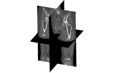
This project addresses the problem of non-invasively estimating the three-dimensional geometry of internal skeletal structures from external body surface scans. In many clinical and biomechanical applications, knowledge of the underlying bone geometry is essential for accurate modeling of musculoskeletal function, assessment of joint loading, and surgical planning. However, obtaining detailed bone geometry traditionally requires medical imaging modalities such as MRI or CT, which are associated with radiation exposure in the case of CT, significant acquisition costs, and limited accessibility in resource-constrained settings. The development of a method that can infer bone geometry directly from non-invasive, low-cost surface measurements would therefore have broad practical implications.
本研究は、外部体表面スキャンから内部骨格構造の三次元形状を非侵襲的に推定する問題に取り組んでいます。筋骨格機能のモデリング・関節負荷の評価・外科的計画の多くの臨床・生体力学応用において、基礎となる骨形状の知識は不可欠です。しかし骨形状の詳細な取得には従来MRIやCTなどの医用撮像モダリティが必要であり、CTの場合は被ばく線量・高い取得コスト・リソースが限られた環境での利用制限という問題があります。非侵襲・低コストの体表面計測から骨形状を推定できる手法は広範な実用的意義を持ちます。
Focus研究焦点
Predicting Internal Body Structures from 3D Surface Scans3D体表面から身体内部の構造を予測
This project addresses the problem of non-invasively estimating the three-dimensional geometry of internal skeletal structures from external body surface scans. In many clinical and biomechanical applications, knowledge of the underlying bone geometry is essential for accurate modeling of musculoskeletal function, assessment of joint loading, and surgical planning. However, obtaining detailed bone geometry traditionally requires medical imaging modalities such as MRI or CT, which are associated with radiation exposure in the case of CT, significant acquisition costs, and limited accessibility in resource-constrained settings. The development of a method that can infer bone geometry directly from non-invasive, low-cost surface measurements would therefore have broad practical implications.
The proposed approach is built around the use of a Statistical Shape Model (SSM), which is a mathematical framework that captures the statistical distribution of shape variations across a population. An SSM is constructed by collecting a large number of 3D bone models derived from medical images of different subjects, aligning them through non-rigid registration, and applying principal component analysis (PCA) to extract the dominant modes of shape variation. The resulting model represents any individual bone shape as the sum of a mean shape and a weighted combination of principal shape components. This compact parameterization significantly constrains the space of plausible bone shapes, making the inference problem substantially more tractable.
The core of the proposed method consists of fitting the SSM to the available external surface data. The 3D surface scan of the body provides information about the external contours of the lower limbs, which are geometrically related to the underlying bone shape through the overlying soft tissue layer. A fitting algorithm is applied to find the SSM parameters that best reconcile the statistical priors of the shape model with the constraints imposed by the external surface observation. This fitting procedure can be formulated as an optimization problem that minimizes the discrepancy between the model-predicted external surface and the observed surface scan, subject to regularization terms that enforce plausible shape configurations.
A key challenge in this approach is the presence of soft tissue between the bone and the external body surface. Soft tissue introduces uncertainty and variability into the relationship between external measurements and bone geometry, as its thickness, distribution, and mechanical properties vary across individuals and anatomical regions. The SSM helps address this challenge by providing strong shape priors that constrain the inference, but the model must also account for inter-individual variability in soft tissue distribution. Future developments may incorporate subject-specific information about soft tissue thickness derived from anthropometric measurements or surrogate measurements to improve accuracy.
The ultimate output of the pipeline is a reconstructed 3D bone model of the lower limbs — including the pelvis, femur, tibia, and fibula — that is geometrically consistent with both the population-level statistics encoded in the SSM and the individual-level morphological constraints provided by the surface scan. This approach has potential applications in subject-specific musculoskeletal modeling for biomechanical research, in clinical settings where rapid assessment of bone geometry is needed without imaging, and as a preprocessing step for other pipelines such as those targeting posture evaluation or bone density analysis.
本研究は、外部体表面スキャンから内部骨格構造の三次元形状を非侵襲的に推定する問題に取り組んでいます。筋骨格機能のモデリング・関節負荷の評価・外科的計画の多くの臨床・生体力学応用において、基礎となる骨形状の知識は不可欠です。しかし骨形状の詳細な取得には従来MRIやCTなどの医用撮像モダリティが必要であり、CTの場合は被ばく線量・高い取得コスト・リソースが限られた環境での利用制限という問題があります。非侵襲・低コストの体表面計測から骨形状を推定できる手法は広範な実用的意義を持ちます。
提案手法は統計的形状モデル(SSM)に基づいています。SSMは集団内の形状変動の統計的分布を捉える数学的フレームワークです。異なる被験者の医用画像から得た多数の三次元骨モデルを収集し、非剛体レジストレーションで位置合わせした後、主成分分析(PCA)を適用して主要な形状変動モードを抽出します。得られるモデルは任意の個人骨形状を平均形状と主成分の重み付き組み合わせとして表現し、この凝縮されたパラメータ化が推定問題を大幅に扱いやすくします。
提案手法の中核は、利用可能な外部体表面データへのSSMのフィッティングです。三次元体表面スキャンは下肢の外形輪郭情報を提供し、この情報は軟部組織層を介して基礎骨形状と幾何学的に関連しています。フィッティングアルゴリズムが形状モデルの統計的事前情報と外部体表面観察の制約を最良に整合させるSSMパラメータを探索します。この手順は、モデル予測外表面と観測スキャンの不一致を最小化しつつ妥当な形状配置を強制する正則化項を含む最適化問題として定式化できます。
本手法の主要な課題は骨と外部体表面の間の軟部組織の存在です。軟部組織の厚さ・分布・機械的特性は個人や解剖学的部位によって異なるため、外部計測と骨形状の関係に不確実性と変動をもたらします。SSMは推定を制約する強力な形状事前情報を提供しますが、軟部組織分布の個人間変動にも対応する必要があります。将来の発展として、精度向上のために人体計測値や代替計測値から得た被験者固有の軟部組織厚情報を組み込むことが考えられます。
パイプラインの最終出力は、SSMにエンコードされた集団レベルの統計的特性と体表面スキャンによる個人レベルの形態学的制約の両方と幾何学的に整合した下肢三次元骨モデル(骨盤・大腿骨・脛骨・腓骨を含む)です。この手法は生体力学研究の被験者固有筋骨格モデリング、撮像なしに迅速な骨形状評価が必要な臨床現場、姿勢評価や骨密度解析などの他パイプラインの前処理ステップとして幅広い応用が期待されます。
Project 9
Sex and Age-related Analysis of Cortical and Trabecular Bone Density in the Femur and Lumbar Spine Using a Large-scale CT Database
大規模CTデータベースを用いた大腿骨と腰椎における皮質骨・海綿骨密度の性別・年代別解析
Bone Mineral Density
Sex-stratified Analysis
CT Analysis
Osteoporosis
Population Study