Daisuke Daisuke
Pelvic Tilt Estimation Using Depth Maps from a Single Plain Radiograph一方向単純X線画像からの深度画像を用いた骨盤傾斜角推定
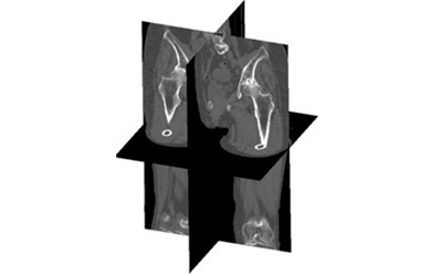
This project focuses on the estimation of pelvic tilt angles — sagittal, coronal, and axial — from a single anteroposterior plain radiograph, using a deep learning-based approach that relies on intermediate depth map representations. Pelvic tilt is a clinically critical parameter in orthopedic practice, particularly in the preoperative planning for total hip arthroplasty (THA). The orientation of the pelvis in three-dimensional space directly influences the functional positioning of the acetabular cup, and inaccurate tilt estimation can lead to suboptimal implant placement, contributing to complications such as dislocation, impingement, or accelerated wear. Currently, the gold standard for measuring pelvic tilt is three-dimensional CT imaging, which provides accurate volumetric data but involves higher radiation doses, greater cost, and longer acquisition times compared to plain radiography.
本研究は、深層学習に基づく深度マップ推定手法を用いて、単一の正面単純X線画像から骨盤の傾斜角(矢状面・冠状面・軸位面)を推定することを目的としています。骨盤傾斜は整形外科臨床において非常に重要なパラメータであり、特に人工股関節全置換術(THA)の術前計画において中心的な役割を果たします。骨盤の三次元的な向きは臼蓋カップの機能的位置決めに直接影響し、傾斜の推定誤差は脱臼・インピンジメント・摩耗促進などの合併症につながるリスクがあります。現在の標準的手法はCT撮影ですが、被ばく線量・費用・撮影時間の面で単純X線に劣ります。
Focus研究焦点
Pelvic Tilt Estimation Using Depth Maps from a Single Plain Radiograph一方向単純X線画像からの深度画像を用いた骨盤傾斜角推定
This project focuses on the estimation of pelvic tilt angles — sagittal, coronal, and axial — from a single anteroposterior plain radiograph, using a deep learning-based approach that relies on intermediate depth map representations. Pelvic tilt is a clinically critical parameter in orthopedic practice, particularly in the preoperative planning for total hip arthroplasty (THA). The orientation of the pelvis in three-dimensional space directly influences the functional positioning of the acetabular cup, and inaccurate tilt estimation can lead to suboptimal implant placement, contributing to complications such as dislocation, impingement, or accelerated wear. Currently, the gold standard for measuring pelvic tilt is three-dimensional CT imaging, which provides accurate volumetric data but involves higher radiation doses, greater cost, and longer acquisition times compared to plain radiography.
The proposed method aims to bridge this gap by enabling CT-equivalent measurements of pelvic tilt directly from low-dose, single-view X-ray images. The core idea is to decompose the problem into two sub-tasks: first, estimating a depth map of the pelvis from the X-ray image; and second, detecting anatomical landmarks whose three-dimensional coordinates can then be used to compute the tilt angles. The two primary landmarks considered in this framework are the anterior superior iliac spines (ASIS) and the pubic tubercle (PT), which together define the pelvic reference plane used in clinical measurement conventions.
To generate the depth maps, a convolutional neural network is trained to regress, for each pixel of the input X-ray, its corresponding depth value relative to the detector plane. This network takes the radiograph as input and predicts a depth image that encodes the three-dimensional distance of the pelvic surface at each image location. The training data for this network is derived from CT volumes, using the same CT-to-X-ray registration pipeline described in related projects: CT scans are aligned to the X-ray coordinate system, digitally reconstructed radiographs are synthesized, and ground-truth depth maps are computed by back-projecting the pelvic surface into the X-ray imaging geometry.
In parallel, a separate landmark detection network predicts the 2D positions of the ASIS and PT landmarks directly in the radiograph. By combining the detected 2D pixel positions with the estimated depth values at those locations, the system reconstructs the full 3D coordinates of each landmark. Once the three-dimensional positions of the landmarks are known, the pelvic tilt angles in all three planes — sagittal, coronal, and axial — can be computed analytically from standard geometric formulas, mirroring the measurement conventions used in CT-based assessment.
The main clinical contribution of this approach is that it enables quantitative, three-dimensional pelvic tilt assessment from a modality that is widely available, inexpensive, and associated with minimal radiation exposure. This could substantially expand access to accurate preoperative planning for THA in settings where CT is not routinely available. The key technical challenge lies in the accuracy of both the depth estimation and the landmark localization, as errors in either component propagate directly into the final tilt angle estimates. Future work will focus on validating the method against CT-derived reference measurements in clinical datasets and on improving robustness to variations in patient positioning and imaging conditions.
本研究は、深層学習に基づく深度マップ推定手法を用いて、単一の正面単純X線画像から骨盤の傾斜角(矢状面・冠状面・軸位面)を推定することを目的としています。骨盤傾斜は整形外科臨床において非常に重要なパラメータであり、特に人工股関節全置換術(THA)の術前計画において中心的な役割を果たします。骨盤の三次元的な向きは臼蓋カップの機能的位置決めに直接影響し、傾斜の推定誤差は脱臼・インピンジメント・摩耗促進などの合併症につながるリスクがあります。現在の標準的手法はCT撮影ですが、被ばく線量・費用・撮影時間の面で単純X線に劣ります。
提案手法はこのギャップを埋め、低被ばく線量の単一方向X線画像からCTと同等の骨盤傾斜計測を実現することを目指しています。問題を二つのサブタスクに分解しています。①X線画像から骨盤の深度マップを推定すること、②解剖学的ランドマークを検出し、その三次元座標から傾斜角を計算することです。主要ランドマークは上前腸骨棘(ASIS)と恥骨結合(PT)であり、これらが臨床的な骨盤基準面を定義します。
深度マップ生成には、入力X線画像の各ピクセルに対してデテクタ平面からの奥行き値を回帰するCNNを使用します。学習データはCT体積データから生成され、CT-X線位置合わせパイプラインを通じてDRRと正解深度マップが算出されます。
並行して、ランドマーク検出ネットワークがX線画像上でASISとPTの二次元座標を予測します。検出された2D座標と推定深度値を組み合わせることで、各ランドマークの完全な三次元座標が復元されます。ランドマークの三次元位置が得られれば、三平面すべての骨盤傾斜角を標準的な幾何学的計算式によって求めることができます。
本手法の臨床的貢献は、低被ばく線量かつ広く利用可能なX線撮影から定量的な三次元骨盤傾斜評価を可能にする点にあります。これにより、CTが日常的に利用できない環境でのTHA術前計画の精度向上と普及が期待されます。今後の課題は、臨床データセットにおけるCT参照値との比較検証と、患者の体位変化や撮影条件の変動に対するロバスト性の向上です。
Project 3
Predicting Survival in Fulminant Myocarditis Using a Large-Scale Pathology Foundation Model
大規模病理基盤モデルを用いた劇症型心筋炎の生存予測
Foundation Model
Survival Prediction
Multimodal Learning
Computational Pathology
Cardiac Imaging