Mamoru Mamoru
Development of an Automated Diagnostic System for Osteonecrosis of the Femoral Head Classification Based on MRI ImagesMRI画像から複数種類の病型分類に基づく大腿骨頭壊死症診断を行う自動システムの構築
This project targets the automated classification of osteonecrosis of the femoral head (ONFH), a debilitating orthopedic condition in which the blood supply to the femoral head is disrupted, leading to ischemic necrosis of the bone. ONFH can progress to femoral head collapse and secondary osteoarthritis if not treated at an early stage, making accurate severity assessment a critical component of clinical management. Providing optimal treatment — ranging from conservative management for mild stages to surgical interventions such as core decompression or total hip arthroplasty for advanced stages — depends fundamentally on an accurate and consistent classification of disease severity. However, current clinical practice faces a significant limitation: even among experienced clinicians, the classification of ONFH severity can be inconsistent, leading to variability in treatment decisions.
本研究は、大腿骨頭壊死症(ONFH)の自動分類システムの開発を目的としています。ONFHは大腿骨頭への血液供給が遮断され骨の虚血性壊死が生じる整形外科疾患であり、早期治療なしには大腿骨頭の圧潰と続発性変形性股関節症へと進行します。軽症への保存療法から進行例への骨頭温存手術・人工関節置換術まで、最適な治療の提供には正確かつ一貫した重症度分類が不可欠です。しかし現状では、経験豊富な臨床医の間でも重症度分類に一貫性が欠けるという課題があります。
Focus研究焦点
Development of an Automated Diagnostic System for Osteonecrosis of the Femoral Head Classification Based on MRI ImagesMRI画像から複数種類の病型分類に基づく大腿骨頭壊死症診断を行う自動システムの構築
This project targets the automated classification of osteonecrosis of the femoral head (ONFH), a debilitating orthopedic condition in which the blood supply to the femoral head is disrupted, leading to ischemic necrosis of the bone. ONFH can progress to femoral head collapse and secondary osteoarthritis if not treated at an early stage, making accurate severity assessment a critical component of clinical management. Providing optimal treatment — ranging from conservative management for mild stages to surgical interventions such as core decompression or total hip arthroplasty for advanced stages — depends fundamentally on an accurate and consistent classification of disease severity. However, current clinical practice faces a significant limitation: even among experienced clinicians, the classification of ONFH severity can be inconsistent, leading to variability in treatment decisions.
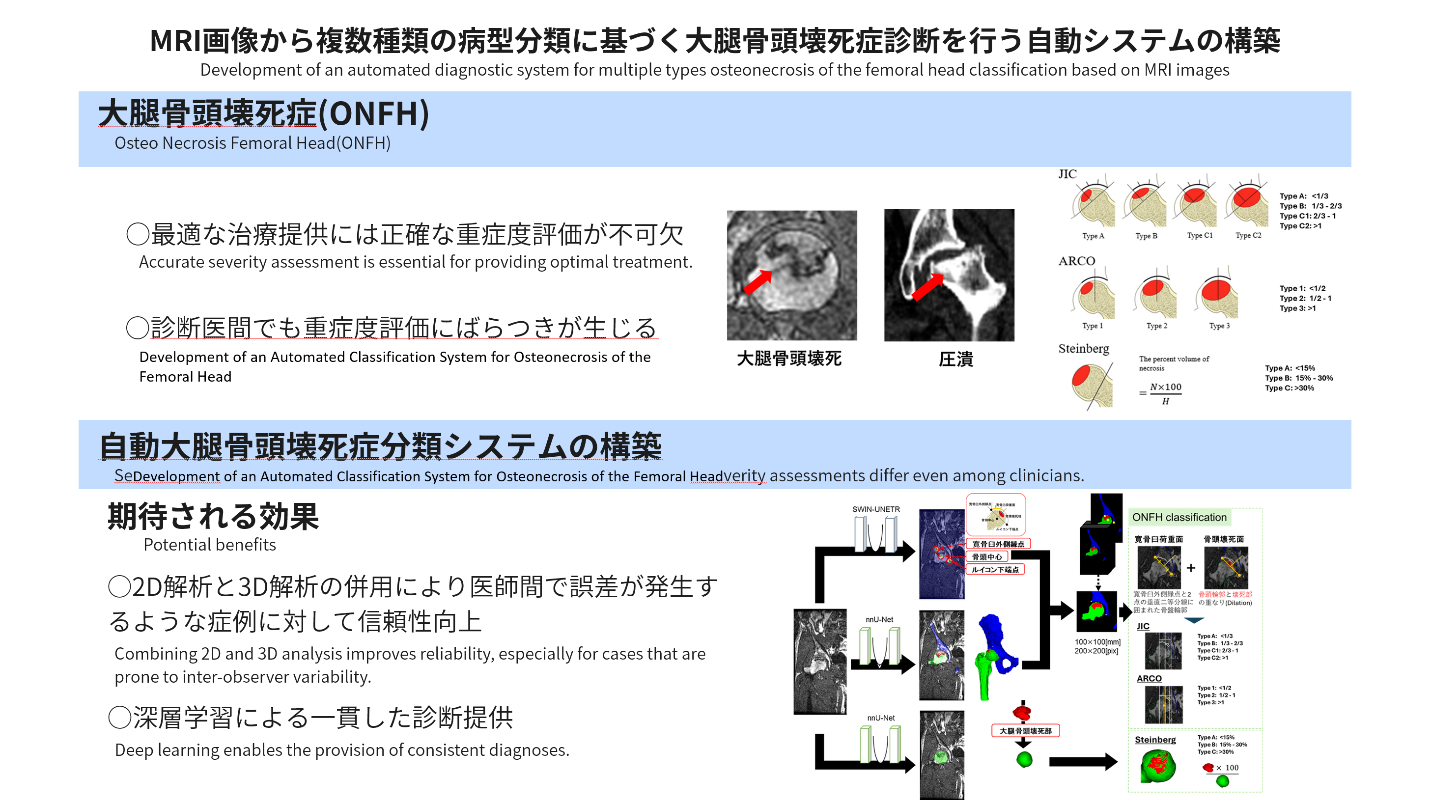
The project addresses this challenge by developing an automated deep learning-based system that can perform ONFH classification according to three major international classification schemes: JIC (Japanese Investigation Committee), ARCO (Association Research Circulation Osseous), and Steinberg. Each of these systems categorizes ONFH using different criteria and thresholds: JIC focuses on the location of the necrotic lesion relative to the acetabulum; ARCO uses a multi-stage scheme based on the extent and structural integrity of the necrotic zone; and Steinberg quantifies the volume of necrosis as a percentage of the total femoral head. Providing simultaneous classification under all three schemes is clinically valuable, as different institutions and countries may use different standards.
The proposed pipeline processes MRI volumes through a cascade of specialized deep learning models. The first stage uses a SWIN-UNETR network to segment the femoral head and identify key anatomical landmarks, including the acetabular rim outer edge, the femoral head center, and the lesser trochanter. These landmarks are essential for establishing the coordinate reference frame used in subsequent analysis. The second and third stages involve two separate nnU-Net models that segment different components of the pathological anatomy: one targeting the necrotic lesion and surrounding transitional zones, and another specifically delineating the femoral head collapse region.
The combination of 2D and 3D analysis is a deliberate design choice motivated by the fact that some classification criteria are best evaluated in specific anatomical planes. For instance, JIC classification requires assessment of the necrotic zone relative to the weight-bearing surface in specific projection views, while Steinberg classification requires accurate volumetric measurement of the necrotic tissue. By integrating both 2D planar information and 3D volumetric information, the system is able to handle cases that are prone to inter-observer variability more reliably than approaches relying on a single analysis modality.
The expected clinical benefits are twofold. First, the system provides consistent, reproducible diagnoses by eliminating the subjective variability introduced by human assessment. Second, it enables simultaneous classification under multiple schemes without additional manual effort, facilitating inter-institutional comparisons and clinical research. The ultimate objective is to deploy this system as a decision-support tool in orthopedic departments, where it can assist clinicians in making timely and evidence-based treatment decisions for ONFH patients.
本研究は、大腿骨頭壊死症(ONFH)の自動分類システムの開発を目的としています。ONFHは大腿骨頭への血液供給が遮断され骨の虚血性壊死が生じる整形外科疾患であり、早期治療なしには大腿骨頭の圧潰と続発性変形性股関節症へと進行します。軽症への保存療法から進行例への骨頭温存手術・人工関節置換術まで、最適な治療の提供には正確かつ一貫した重症度分類が不可欠です。しかし現状では、経験豊富な臨床医の間でも重症度分類に一貫性が欠けるという課題があります。
本研究はこの課題に対し、国際的な三大分類体系(JIC・ARCO・Steinberg)に基づいてONFH分類を実行できる深層学習自動システムを開発することで取り組みます。JICは臼蓋に対する壊死病変の位置を基準とし、ARCOは壊死帯の範囲と構造的完全性に基づく多段階スキームを使用し、SteinbergはMRI上の壊死体積を大腿骨頭全体のパーセンテージで定量化します。三種の分類体系を同時に提供することは、施設や国によって使用基準が異なる臨床現場において高い価値を持ちます。
提案パイプラインは、特化した深層学習モデルのカスケードを通じてMRIボリュームを処理します。第一段階ではSWIN-UNETRネットワークが大腿骨頭をセグメント化し、臼蓋外縁・骨頭中心・小転子などの主要ランドマークを同定します。第二・第三段階ではそれぞれ異なるnnU-Netモデルが壊死病変の周囲領域と骨頭圧潰部位をセグメント化します。
2D・3D解析の組み合わせは、分類基準によって特定の解剖学的断面での評価が最適となる場合があるという事実に基づく意図的な設計です。JIC分類は特定の投影ビューで荷重面に対する壊死帯の評価を必要とし、Steinberg分類は壊死組織の正確な体積計測を必要とします。2D平面情報と3D体積情報の統合により、観察者間変動が生じやすい症例においても単一解析モダリティより信頼性の高い診断が実現します。
期待される臨床的効果は二つあります。第一に、人間の評価による主観的変動を排除した一貫性・再現性のある診断の提供。第二に、追加の手動作業なしに複数分類体系での同時分類を可能にし、施設間比較と臨床研究を促進することです。最終目標は、整形外科部門での意思決定支援ツールとしてのシステム導入により、ONFH患者への適切な根拠に基づく治療決定を支援することです。
Project 6
Anterior-Posterior Depth Estimation from a Single X-ray Image
単一方向X線画像からの前後面深度推定
Depth Map Estimation
3D Reconstruction
Vertebral Analysis
X-ray Imaging
2D-3D Registration